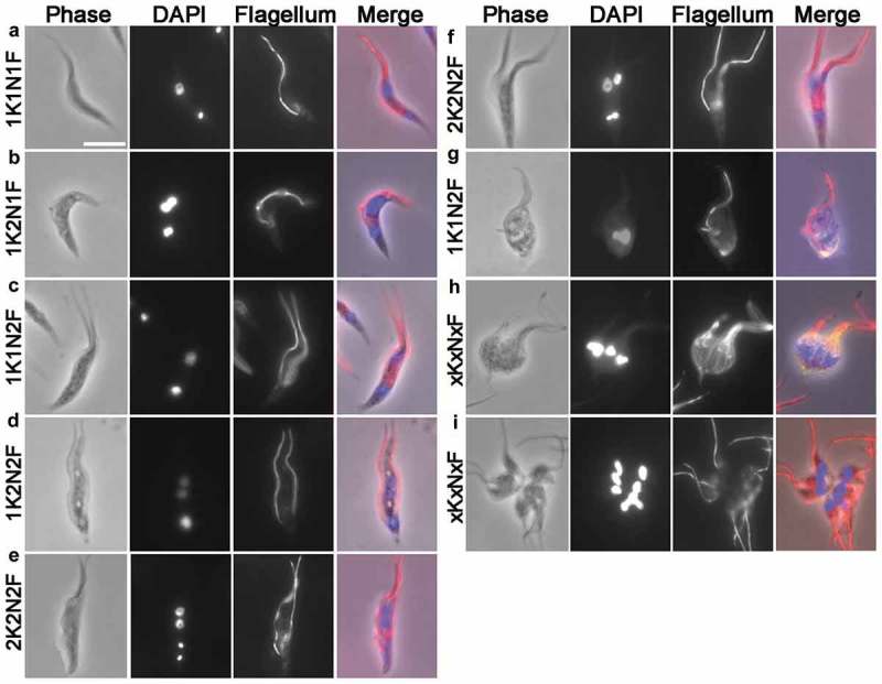
Figure 7.

Immunofluorescence staining of T. lewisi cells using anti-paraflagella rod antibodies. Trypanosomes were settled onto glass microscope slides, fixed in methanol at −20°C, and a mouse monoclonal antibody (mAb) against the T. brucei flagellum L8C4 was used as primary antibody, followed by secondary antibody (goat anti-mouse IgG, Invitrogen). DNA was stained by DAPI and cells were categorized according to their number of nuclei (N), kinetoplasts (K) and flagella (F). (a) 1K1N1F, (b) 1K2N1F, (c) 1K1N2F, (d) 1K2N2F, (e and f) 2K2N2F, (g) 1K1N2F (trypomastigote form in transition towards a dividing epimastigote), (h,i) xKxNxF. In the merged images, the red staining represents the flagellum while the blue staining highlights the nuclei and kinetoplasts. Scale bar represents 10 μm.
