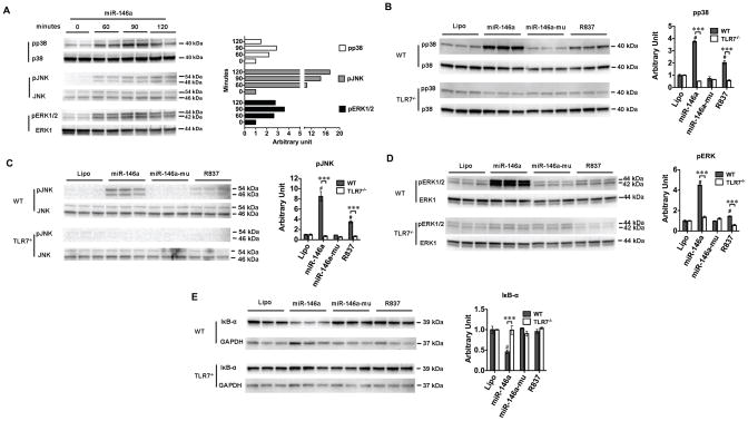
Fig. 6. miR-146a activates MAPK and NF-κB signaling via TLR7.
A. Immunoblotting of p38, JNK and ERK1/2. Macrophages were treated with 50 nM of miR-146a. At time 0, 60, 90, 120 min, cells were lysed and tested for phosphorylated and total p38, JNK, and ERK1/2. The difference was expressed as the ratio to time 0 as quantified by Image J. B–D. TLR7 deletion completely blocked miR-146a- or R837-induced phosphorylation of p38, JNK, and ERK1/2, respectively. WT and TLR7−/− macrophages were treated with miR-146a (50 nM) or R837 (0.25 μg/ml) for 90 minutes. Cell lysate extracted and analyzed for phosphorylation of p38, JNK and ERK1/2. Total MAP kinase expression was served as the internal control. # P < 0.001 vs. lipofectamine control (Lipo). *** P < 0.001, WT vs. TLR7−/− group. n=3 in each group. The difference was expressed as the ratio to lipofectamine control (Lipo) in each strain as quantified by Image J. E. TLR7 deletion blocked miR-146a-induced IκB-α degradation. GAPDH served as the protein loading control. # P < 0.001 vs. lipofectamine control (Lipo). *** P < 0.001, WT vs. TLR7−/− group. n=3 in each group. The difference was expressed as the ratio to lipofectamine control (Lipo) in each strain as quantified by ImageJ.

