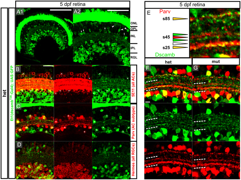
Figure 5 – dscamb is expressed in ACs and RGCs subtypes, but is not required for IPL lamination.
A1) Et1(dscambt2b:Gal4) expression in retinal section from 5dpf larva. Dorsal is left, medial is up. Scale bars: 100μm.
A2) Higher magnification image of A1. Arrowheads: BCs, arrows: ACs. ONL: outer nuclear layer, OPL: outer plexiform layer, INL: inner nuclear layer, IPL: inner plexiform layer, RGL: retinal ganglion cell layer. Scale bar: 50μm.
B-D) Confocal images of IPL in sections from 5dpf heterozygous larvae. Scale bar: 20μm.
B) Et1(dscambt2b:Gal4) compared to pan-AC marker (5E11; red). Asterisks: GFP+/5E11+ ACs.
C) Et1(dscambt2b:Gal4) compared to Parv+ AC marker (red). Asterisks: GFP+/Parv+ ACs.
D) Et1(dscambt2b:Gal4) compared to pan-RGC marker (Hermes; red).
E) s25, s45, and s85 Parv+ sublaminae in the IPL. Triangles indicate sublaminae that are GFP+/Parv+ (yellow), GFP+ only (green), Parv+ only (red), or negative for both (empty). Scale bar: 5um.
F,G) Images of IPL in 5dpf heterozygous (F) and homozygous (G) dscamb mutant larvae. White dashed lines outline region containing s25 and s45 Parv+ sublaminae. Scale bar: 5um.

