
Figure 1.
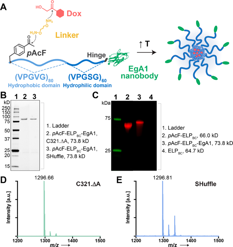
Design and expression of pAcF-ELPBC-EgA1. (A) Schematic showing design and assembly of Dox-pAcF-ELPBC-EgA1 nanoparticles. Dox (red) is conjugated to the pAcF residue (dark gray) at the N-terminus of the amphiphilic ELPBC chain (blue) by a telechelic hydroxylamine linker (yellow). The hydrophilic ELP block is fused to the EgA1 nanobody (green) by a flexible hinge (black). (B) SDS-PAGE of pAcF-ELPBC-EgA1 in C321.ΔA (lane 2) and SHuffle E. coli (lane 3). (C) Fluorescence imaging of SDS-PAGE qualitatively confirms reactivity of the ketone group on the pAcF residue in pAcF-ELPBC (lane 2) and pAcF-ELPBC-EgA1 (lane 3) with a hydroxylamine dye. ELPBC without pAcF incorporated is not labeled (lane 4). Tryptic digest of these constructs followed by MALDI-TOF-MS shows a single peak that is consistent with the incorporation of a pAcF residue in pAcF-ELPBC-EgA1 of protein expressed in (D) C321.ΔA (1296.66 m/z) and (E) SHuffle E. coli (1296.81 m/z).