
Figure 2.
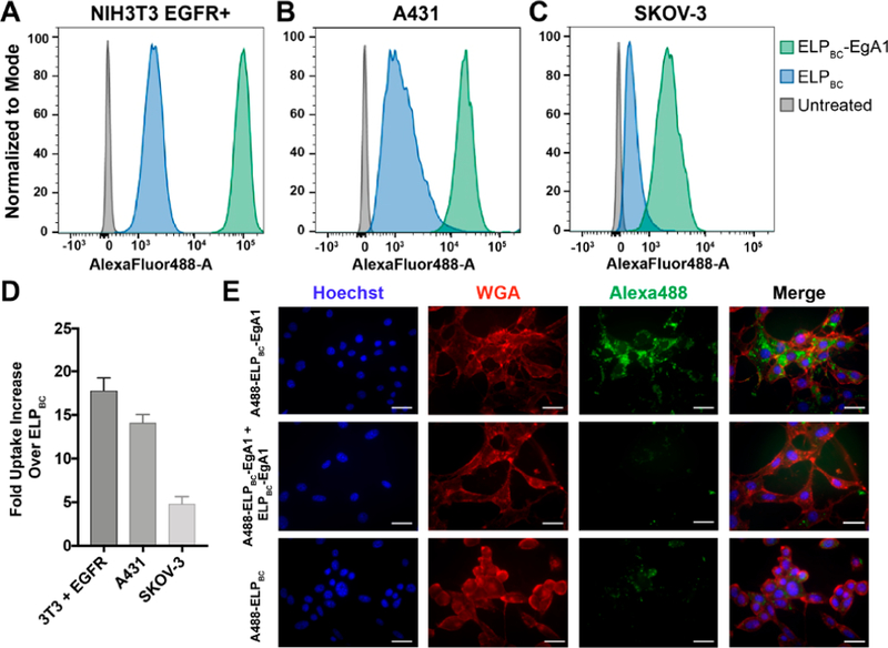
Flow cytometry confirms specificity of the EgA1 nanobody for human epidermal growth factor receptor (EGFR). Fluorescently labeled ELPBC-EgA1 or ELPBC were incubated with various cell lines and analyzed by flow cytometry. (A) NIH3T3 murine fibroblasts transfected with human EGFR (NIH3T3 EGFR+); (B) A431 squamous carcinoma cells; and (C) SKOV-3 ovarian adenocarcinoma cells all indicate enhanced uptake of the ELPBC-EgA1 as compared to ELPBC. (D) Geometric mean fluorescent intensities (gMFI) of the cell populations were used to quantify the fold uptake of ELPBC-EgA1 over ELPBC and shows the range of nanobody-mediated targeting of EGFR across the cell lines. (E) Fluorescence microscopy images of NIH3T3 EGFR+ cells incubated with AlexaFluor488-ELPBC-EgA1, AlexaFluor488-ELPBC-EgA1 with 10-fold excess unlabeled ELPBC-EgA1, and AlexaFluor488-ELPBC shows increased uptake of the AlexaFluor488-ELPBC-EgA1 construct, while the cells incubated with 10-fold excess unlabeled ELPBC-EgA1 demonstrate the specificity of the EgA1 nanobody. The samples incubated with AlexaFluor488-ELPBC show the basal level of nanoparticle uptake. Nuclei stained with Hoechst (blue), cell membranes stained with AlexaFluor594-wheat germ agglutinin (red), and all ELP constructs with AlexaFluor488 (green). Scale bars 25 μm.