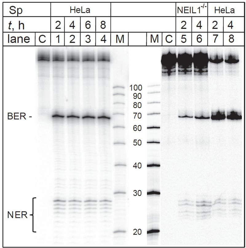
Figure 5.
The spiroiminodihydantoin lesions are substrates of BER and NER in intact cells. The 32P-internally labeled DNA hairpins harbouring Sp lesions were transfected either into fully BER/NER-competent HeLa cells (lanes 1–4), or into NEIL1−/− cells that lack the glycosylase NEIIL1 (lanes 5–8) that is known to excise Sp lesions in double-stranded DNA.19, 20 The incubation times (t) varied between 2 and 8 hours. Lanes M: oligonucleotide size markers (We note that the intensity of the synthetic maker Lane M only, was diminished separately from the rest of the gel because it was oversaturated). Lanes C: Control experiments; the hairpins were not transfected into the cells, but were subjected to the same treatment otherwise. The panel shown is a composite of autoradiographs of two gels.

