
Figure 1.
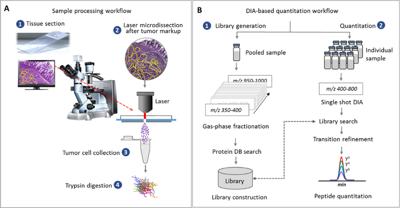
(A) Proteomic analysis of FFPE biopsy samples. Tissue sections mounted on Director® slides were subjected to microdissection (10 μm tissue thickness) after tumor-specific markup. Collected tumor cells were proteolyzed by trypsin after heat treatment. (B) DIA analysis for quantitative analysis. A library containing chromatographic and mass spectrometric attributes (MS1 and MS2 traces) of detected peptides was constructed using the gas-phase-fractionated DIAs in a pooled sample with 2 Th m/z window18. This library was used to match peptides from the DIA data obtained from the individual sample using a wider isolation window (20 Th). Summed AUCs of the fragment ions’ chromatograms per precursor are used to generate quantitative data.