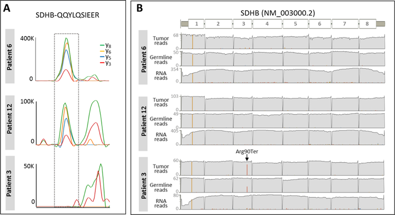
Figure 4.

Differential SDHB status. (A) Chromatographic traces of four extracted fragment ions (y3, y5, y6, y8) (± 10 ppm) of a unique SDHB peptide, QQYLQSIEER are shown for three biopsies exhibiting different levels (high, intermediate, absent) of SDHB protein. (B) Depth of tumor DNA, germline DNA, and tumor RNA sequencing reads mapped to SDHB in three biopsies corresponding to the patient samples shown in DIA-MS data left. Red bar indicates the number of reads that contain Arg90ter.
