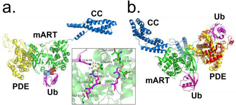
Figure 2.
Ubiquitin recognition by the SidE ligase. (A) Structure of SidE in complex with ubiquitin (Ub) (magenta) at mART site with key interacting residues of Ub highlighted. The hydrophobic patch of Ub is depicted in cyan, and key Arg residues depicted in red. Inset depicts polar/charge interactions between the SidE mART domain and Ub. (B) Structure of SdeA in complex with Ub at mART site (magenta) aligned with structure of PDE domain of SdeD (red) in complex with ADPR-Ub (magenta). The SdeD L.p. effector has a domain homologous to the SidE PDE domain with the same catalytic residues but lacks an mART domain. Abbreviations: L.p., Legionella pneumophila; mART, mono-ADP ribosyltransferase; PDE, phosphodiesterase; ADPR-Ub, ADP ribosylated ubiquitin.

