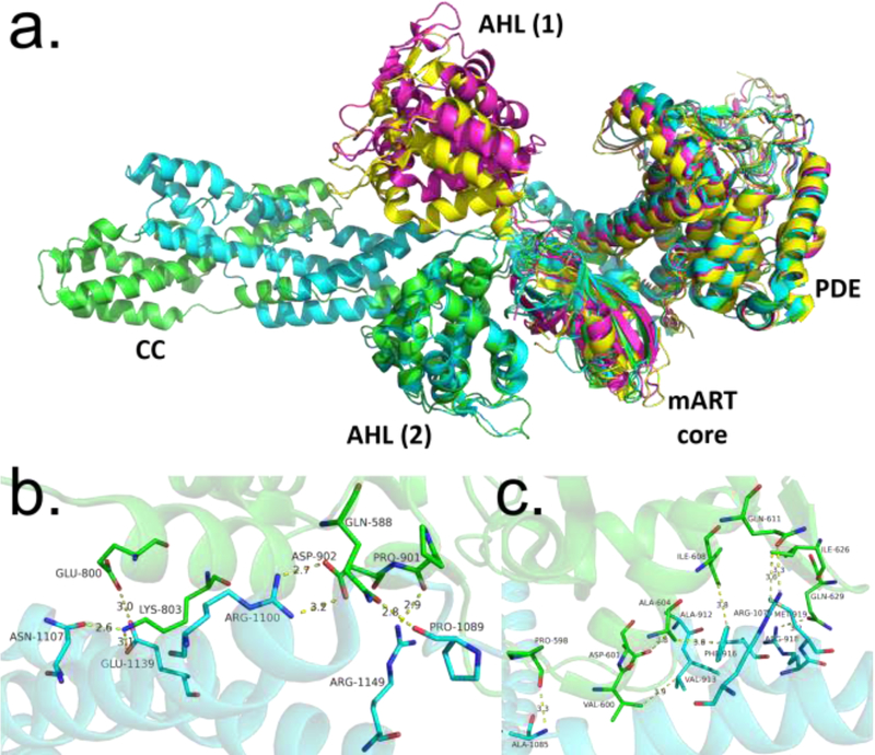
Figure 4.
Comparison of SidE and SdeA structures. (A) Structural alignment of various constructs of SidE proteins (green: SidE222−1057, cyan: SdeA231−1190, magenta: SdeA211−910, yellow: SdeA213–907), with domains labeled. AHL (1) and AHL (2) refer to two different orientations of the AHL domain observed in crystal structures of the SidE constructs, with the latter corresponding to a productive orientation. (B) Interactions between mART core and CC domain of SdeA. (C) Interactions between mART AHL and CC domain. Abbreviations: AHL, α-helical lobe; CC, coiled-coil; mART, mono-ADP ribosyltransferase.

