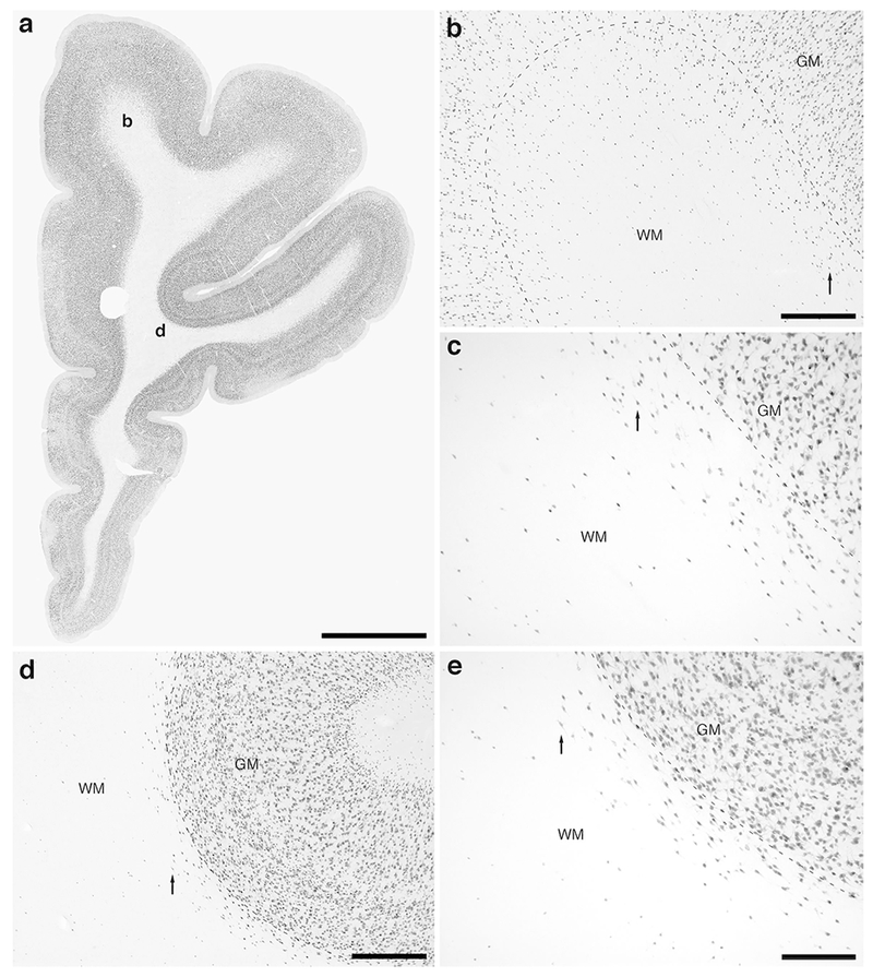
Figure 1:

Photomicrographs of neuronal nuclear marker (NeuN) immunostaining in a coronal section through the rostral portion of the frontal lobe of the lar gibbon, deep to the granular prefrontal cortex, showing the distribution of infracortical white matter interstitial cells (WMICs), presumably neurons. (a) Low magnification image of the entire section through the right frontal lobe of the lar gibbon stained with NeuN, showing the presence of numerous cells deep to the cortex. (b) Moderately magnified image of the superior frontal gyrus (from the region indicated by the b in image a), showing the numerous WMICs deep to the cerebral cortex (grey matter, GM) within the infracortical white matter (WM). The approximate boundary of the deep border of cortical layer VI and the WM is marked by a dashed line. (c) High magnification image of the cortical/white matter boundary (marked by a dashed line) of the superior frontal gyrus, showing the WMICs deep to the cerebral cortex within the WM. Arrows in b and c indicate the same neuron for orientation of image location. (d) Moderately magnified image of the fundus of the inferior frontal gyrus (from the region indicated by the d in image a), showing WMICs deep to the cerebral cortex (GM) within the WM. The approximate boundary of the deep border of cortical layer VI and the WM is marked by a dashed line. (e) High magnification image of the cortical/white matter boundary (marked by a dashed line) of the fundus of the inferior frontal gyrus, showing the WMICs deep to the cerebral cortex within the WM. Arrows in d and e indicate the same neuron for orientation of image location. Scale bar in a = 5 mm and applies to a only. Scale bars in b and d = 500 μm and apply to b and d only. Scale bar in e = 250 μm and applies to c and e. In all images dorsal is to the top of the image and medial to the left.
