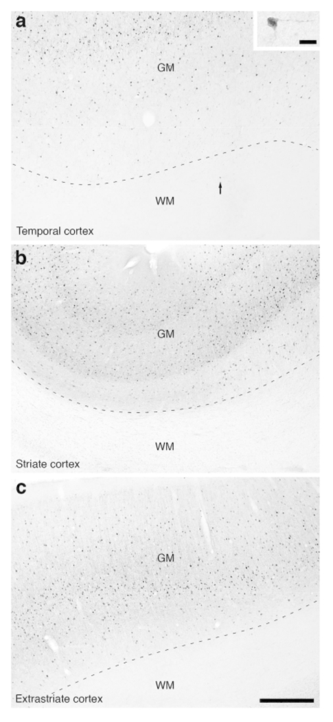
Figure 16:

Photomicrographs of PV immunostaining in the white matter of the lar gibbon, showing the distribution of occasional WMICs immunoreactive to PV. (a) White matter (WM) deep to the temporal cortex, with an arrow indicating a single PV-immunoreactive WMIC. The inset in a shows this solitary stained neuron at a higher magnification. (b) PV-immunoreactive WMICs were not observed in the white matter deep to the striate cortex. (c) PV-immunoreactive WMICs were not observed in the deep white matter surrounding the extrastriate cortex. Scale bar in c = 500 μm and applies to a, b and c. Scale bar in inset of a = 20 μm and applies to the inset only. In all images dorsal is to the top of the image and medial to the left.
