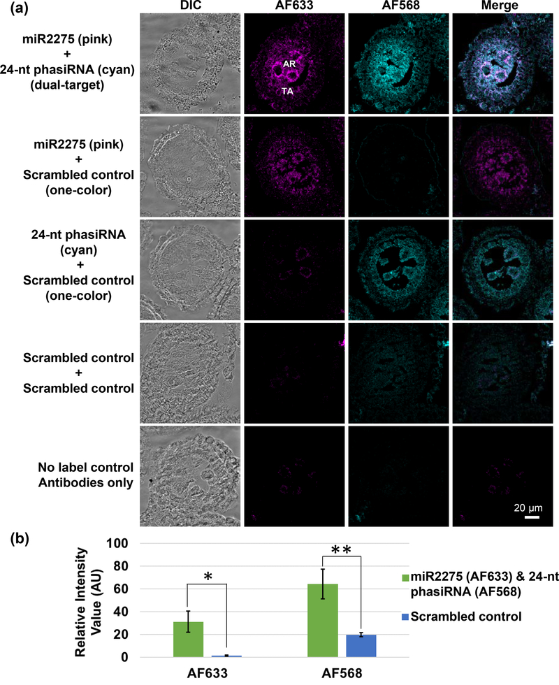
Figure 3. Dual-target sRNA-FISH for maize anthers.
(a) sRNA-FISH detected both zma-miR2275 (detected in the AF633 channel; magenta) and the 24 nt phasiRNA (detected in the AF568 channel; cyan) in the tapetal layer and archesporial cells. Each image was collected in spectra mode with laser scanning confocal microscopy and then spectrally unmixed using Zen Software. Bright-field and merged images were also shown for each image. TA, tapetal layer; AR, archesporial cells. Scale bars = 20 μm for all images. (b) Quantification of the AF633 and AF568 signal intensity in dual-target sRNA FISH and controls. (Significance level: < 0.05, *; < 0.01, **).

