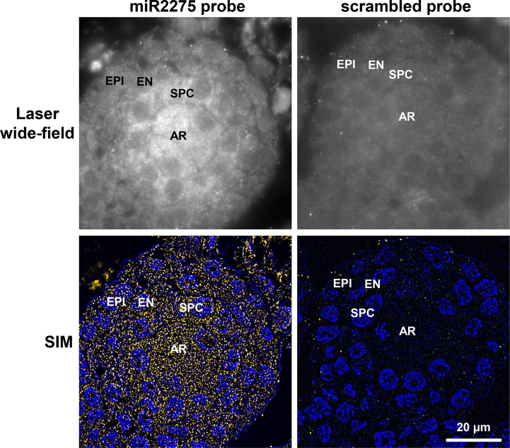
Figure 5. Localization of miR2275 in premeiotic maize anthers using SIM.
Top left panel: Laser wide-field images shown miR2275 is detected in the archesporial cells and secondary parietal cells; the latter give rise to the middle layer and tapetum. Bottom left panel: detection of miR2275 using super-resolution structured illumination. miR2275 is localized to archesporial and secondary parietal cells. Right panels are images of the scrambled probe control. AR, archesporial cells; SPC, secondary parietal cells; EN, endothecium; EPI, epidermis. Scale bar = 20 μm for all images.

