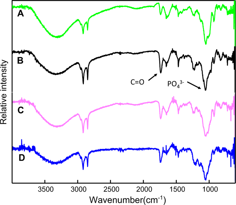
Fig. 3.
FTIR spectra of the minerals obtained from the mineralization assays under incubation of TNAP-proteoliposomes composed of (green) DPPC, (black) DPPC:Chol (9:1), (pink) DPPC:SM (9:1) and (blue) DPPC:Chol:SM (8:1:1) (molar ratios), in SCL, at 37 °C, at pH 7.5, in the absence of nucleators. ATP concentrations of 6 mM, 10 mM, 5 mM, and 9 mM were used for the proteoliposomes composed of DPPC, DPPC:Chol, DPPC:SM and DPPC:Chol:SM, respectively, as indicated by arrows in Fig. 1. Mineralization was followed by the differences in the ratio between the areas of the internal reference band of the phospholipid (C=O) at 1740 cm−1 and the band corresponding to the asymmetrical stretching of the PO 43− group at 1032 cm−1

