Figure 4.

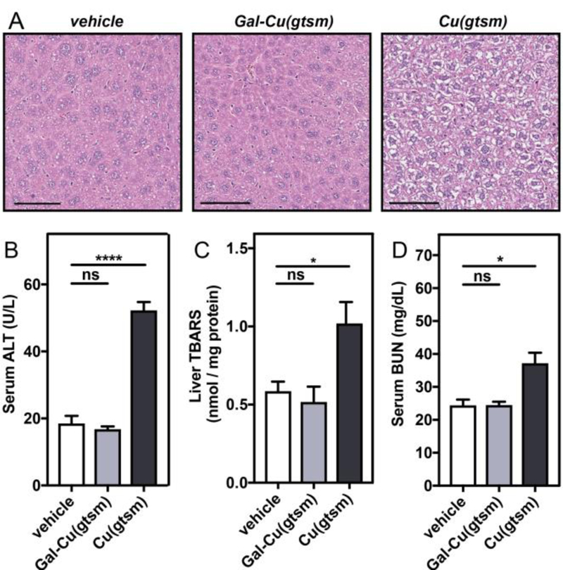
Gal-Cu(gtsm) treatment is non-toxic despite delivering more copper than Cu(gtsm). (A) Representative liver tissue slices from H&E staining show significant hydropic degeneration (wispy/white cytosolic areas surrounding nuclei) upon Cu(gtsm) treatment. Liver sections were isolated six hours following vehicle or 0.75 mg Cu/kg mouse ionophore i.p. injections. Scale bar = 100 µM. (B-D) Toxicity assays were performed on serum (B,D) or liver lysate (C) collected from mice treated with Cu(gtsm) or Gal-Cu(gtsm) at 0.75 mg Cu/kg mouse after 6 hours to evaluate liver (B,C) and kidney (D) toxicity. Error bars = SEM (n = 5). Statistical analyses were performed with a one-way ANOVA with Bonferroni’s multiple comparisons test where *P ≤ 0.05, ****P ≤ 0.0001.
