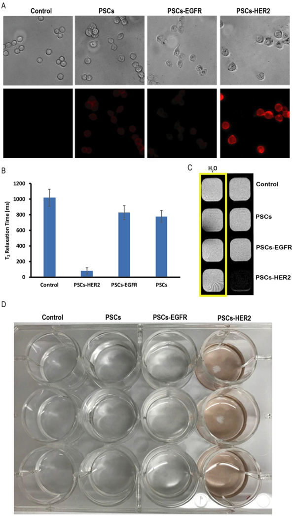
Figure 3.

(A) Phase contrast (top row) and fluorescence microscopy (bottom row) images of HER2/neu-positive T617 cells incubated without nanoemulsions (control) and with PSCs, PSCsEGFR and PSCs-HER2 for 1h. (B) Relaxivity measurements of T617 cells incubated with targeted and non-targeted nanoemulsions. (C) MR phantom image of T617 cells after incubation with targeted and non-targeted PSCs for 1h. (D) Photograph of T617 cells in a 12 well-plate following incubation with targeted and non-targeted PSCs.
