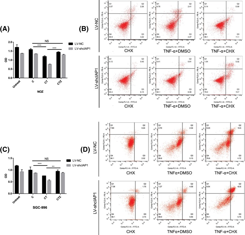
Figure 3. cIAP1 enhances TNF-α/cycloheximide-induced apoptosis in GBC.
(A,C) CCK-8 test at indicated time revealed co-administration of TNF-α (40 ng/ml) and cycloheximide (8 μg/ml) blunt cell viability instead of administration of TNF-α alone and the effect could be retrieved by 1 h pre-treatment by pan-Caspase inhibitor zVAD (20 mM). In each subgroup, the LV-shcIAP1 presented a lower viability and those with treatment of TNF-α and cycloheximide present the least (**P< 0.01, ***P< 0.001). (B,D) Similar groups were subscribed to flow cytometry for analysis of apoptosis. Apoptosis were observed obviously in TNF-α/cycloheximide group and even more ratio of apoptotic cells was observed in LV-shcIAP1 group. All the experiments were performed three times with similar results in both cell lines.

