Figure 2.

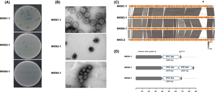
Morphological and molecular characterization of bacteriophages KN1‐1, KN3‐1 and KN4‐1.A. The plaque morphology of phages KN1‐1, KN3‐1 and KN4‐1. Clear plaques surrounded by translucent halos were observed on the individual lawns of Klebsiella A1517 (KN1), N386 (KN3) and 4565 (KN4).B. Electron micrographs of phages KN1‐1, KN3‐1 and KN4‐1. Purified phage particles of KN1‐1, KN3‐1 and KN4‐1 exhibited icosahedral capsids and short tails.C. Genome comparisons of phages KN1‐1, KN3‐1, KN4‐1 and K5‐2. Comparative analysis of the phage genomes was performed with Easyfig. The four phage genomes are very similar except for a 2.5–4.5 kb variable region (indicated by an asterisk).D. The variable region. Genes located in this variable region are shown as white arrows, while the conserved genes upstream and downstream are shown in grey. Gene length is shown in parentheses; the axis below shows the position in kb.
