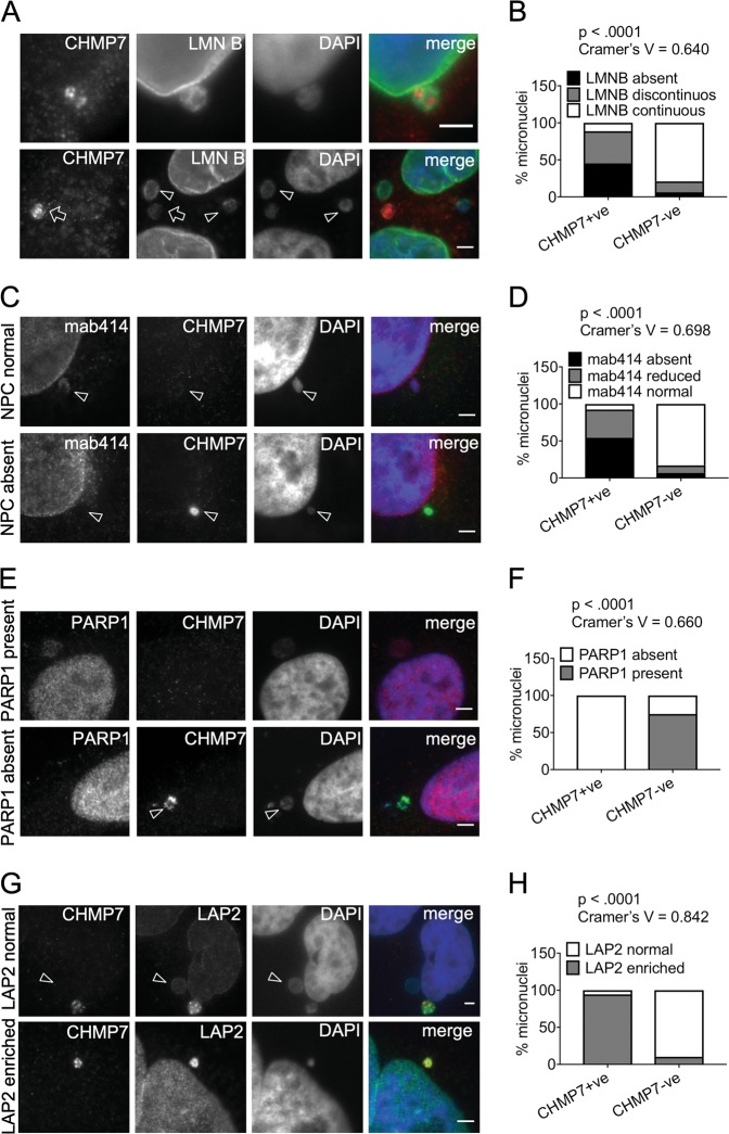
Fig. 4. Micronuclei with CHMP7 accumulations lack nuclear lamina integrity, nuclear pore complexes, and soluble nuclear proteins but are enriched in LAP2.
a Immunofluorescence image of a micronucleus where CHMP7 localizes to regions with gaps in Lamin B in control cells (top panel). Examples of CHMP7-positive (arrows) and negative (arrowheads) in control HeLa cells (bottom panel). Scale bar 3 µm. b Distribution of the status of CHMP7 and Lamin B in micronuclei (minimum 100 micronuclei scored per repeat). c Examples of CHMP7-positive and negative micronuclei with NPC staining in control HeLa cells. d Micronuclei in HeLa cells were scored for the status of CHMP7 and the status of NPC (mab414) within the same micronucleus (minimum 160 micronuclei scored per repeat). e Examples of CHMP7-positive and negative micronuclei showing absence or presence of PARP1 staining. Scale bar 3 µm. f Quantification of the presence or absence of PARP1 and CHMP7 staining within the same micronucleus (minimum 200 micronuclei scored per repeat). g Examples of CHMP7-positive and negative (arrowhead) micronuclei with LAP2 staining. Scale bar 3 µm. h Quantification of the presence or absence of LAP2 and CHMP7 staining within the same micronucleus (300 micronuclei per repeat). Data were analyzed using Fishers’ exact test on pooled raw counts distributions (N = 3)

