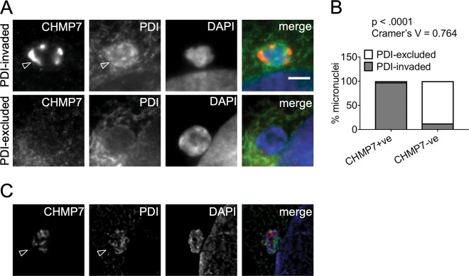
Fig. 5. Micronuclei with ESCRT-III accumulations display ER membrane invasion.
a Examples of CHMP7-positive and negative micronuclei with PDI staining. PDI-excluded micronuclei have endoplasmic reticulum (ER) membrane surrounding their boundaries but not in the micronuclear interior. PDI-invaded micronuclei show ER membrane inside the micronuclear boundary, indicating nuclear envelope collapse (arrowheads). b Distribution of the status of CHMP7 and the presence or absence of PDI invasion within the same micronucleus (minimum 100 micronuclei scored per repeat). Data were analyzed using Fishers’ exact test on pooled raw counts distributions (N = 3). c Example image of a micronucleus in a Hela cell after treatment with VPS4 sRNAi for 48 h. Accumulation of PDI and CHMP7 within the micronucleus and the irregular shape display membrane infiltration (arrowhead). Scale bars 3 µm

