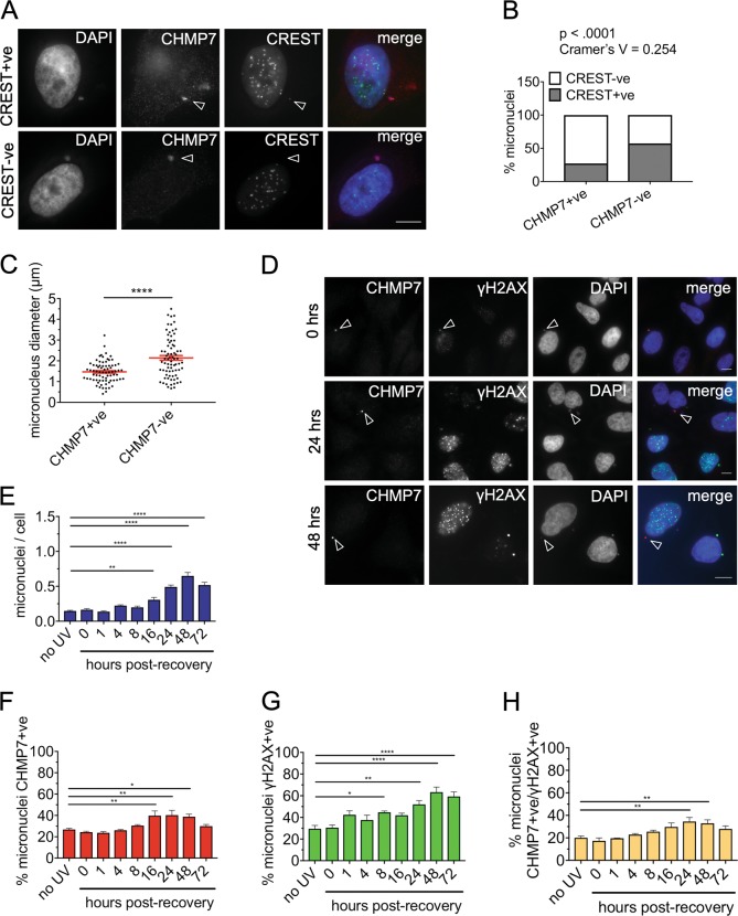
Fig. 6. ESCRT-III preferentially accumulates on acentric micronuclei.
a HeLa cells transfected with either control or VPS4 sRNAi for 48 h hours and co-stained for CREST to detect centromeres, CHMP7, and DAPI. Examples of CHMP7 + ve micronuclei that are CREST + ve (arrowhead indicating weak CREST signal) or CREST-ve (arrowhead indicating the absence of CREST signal). Scale bars 10 µm. b Quantification of micronuclei in HeLa cells containing at least one definite CREST foci which resembles those found in the primary nucleus and CHMP7 (minimum 200 micronuclei scored per repeat). Results were analyzed using a Fishers exact test on pooled data. Individual biological repeats were also significant to Fishers exact (p < 0.05). c Control HeLa cells were stained for CHMP7 and DAPI. The diameter of micronuclei measured in ImageJ (75 CHMP7-positive and 75 CHMP7-negative micronuclei). HeLa cells were transfected with sRNAi for 48 h and stained to show CREST, CHMP7, and DAPI. d–h HeLa cells were treated with a 10 mJ/cm2 dose of UV-C irradiation and fixed at various timepoints following treatment. The cells were stained for γH2AX, CHMP7, and DAPI. d Accumulations of CHMP7 and γH2AX within a single micronucleus in control cells and in cells 24 and 48 h post recovery from UV-C (scale bar 10 µm). The number of micronuclei per cell post UV-C treatment is shown in e. Micronuclei were scored for presence of f CHMP7 accumulation and g γH2AX foci, with micronuclei containing at least one focus being considered as positive (400 cells per treatment). The percentage of micronuclei positive for both γH2AX and CHMP7 are shown in h. The percentage is calculated against the total number of micronuclei counted for each time point, varying between a minimum of 50 and a maximum of 285. Averages and SEM are shown (N = 3). Results were analyzed using a one-way ANOVA with Dunnett’s post hoc test

