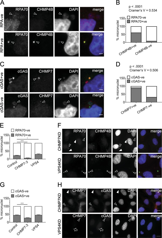
Fig. 7. CHMP7 is important for generating damaged DNA at micronuclei.
a Control HeLa cells containing micronuclei positive or negative for RPA70 and CHMP4B. Top panel: the arrowheads show micronuclei negative for RPA70 and CHMP4B. Bottom panel: the arrowheads show micronuclei positive for RPA70 and CHMP4B. Scale bar 3 µm. b Micronuclei, in control HeLa cells, scored for the status of CHMP7 and for presence or absence of single-stranded DNA (RPA70) within the same micronucleus (minimum 115 micronuclei scored per repeat). Data analyzed using Fishers’ exact test on pooled raw counts distributions (N = 3). c Control HeLa cells with micronuclei positive or negative for cGAS and CHMP7. Top panel: the arrowheads show micronuclei positive for cGAS (red) and CHMP7 (green). Bottom panel: the arrowheads show micronuclei negative for cGAS (green) and CHMP7 (red). Scale bar 3 µm. d Micronuclei, in control HeLa cells, scored for the status of CHMP7 and the presence or absence of cGAS within the same micronucleus (minimum 150 micronuclei scored per repeat). Data analyzed using Fishers’ exact test on pooled raw counts distributions (N = 3). e Micronuclei from HeLa cells transfected for 48 h with the indicated siRNA and scored for enrichment in RPA70 signal (minimum 50 total micronuclei scored per treatment, per repeat). Results were analyzed using a one-way ANOVA with Dunnett’s post hoc test. Averages and SEM shown (N = 3). f Top panel: HeLa cells depleted of CHMP7 for 48 h (arrowheads show RPA70 + ve micronuclei; filled arrowheads show RPA70-ve micronuclei). Bottom panel: HeLa cells depleted of VPS4 for 48 h (arrowheads show RPA70 + ve and CHMP4B + ve micronuclei). Scale bar 10 μm. g Micronuclei in HeLa cells transfected for 48 h with the indicated siRNA and scored for enrichment in cGAS signal (minimum 55 micronuclei scored per treatment, per repeat). Results analyzed using a one-way ANOVA with Dunnett’s post hoc test. Averages and SEM shown (N = 3). h Top panel: HeLa cells depleted of CHMP7 for 48 h (arrowheads show cGAS + ve micronuclei; filled arrowheads show cGAS-ve micronuclei). The CHMP7 channel is 3× over-exposed to highlight the absence of CHMP7 accumulations on the micronuclei. Bottom panel: HeLa cells depleted of VPS4 for 48 h (arrowhead shows a cGAS + ve micronucleus; arrow shows cGAS enrichment at a CHMP7 nuclear accumulation). Scale bar 10 μm. f and h the exposure time has been adjusted to compensate for varying intensity of the signals between CHMP7 and VPS4 knockdown experiments

