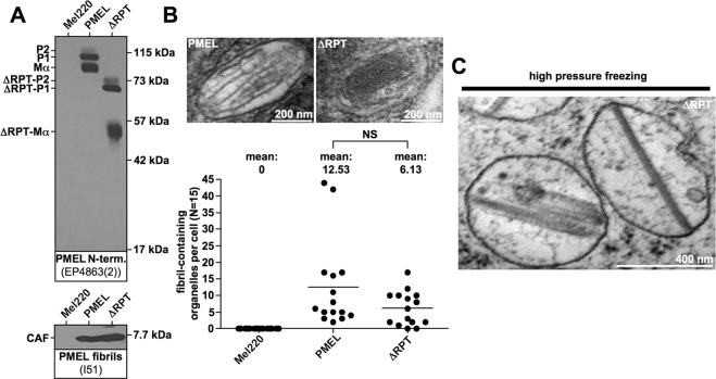
Figure 1.
The RPT domain controls the morphology of human PMEL amyloid. (A) Western blot analysis of SDS-lysed total membranes using PMEL-specific antibodies EP4863(2) (PMEL N-term.) and I51 (CAF). (B) Conventional TEM and quantitative EM analysis of Mel220 transfectants showing the number of fibril-containing organelles per cell [N = 15]. An unpaired two-tailed t-test was used to determine whether means are statistically different from the wt-PMEL sample (NS, not significant). Representative electron micrographs are depicted. (C) EM analysis of ΔRPT-expressing Mel220 cells prepared by high pressure freezing and freeze substitution.

