Fig. 5.
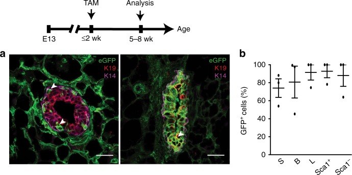
PDGFRα+ cell descendents contribute to early mammary epithelial development. a Immunofluorescent images of GFP-labeled cells with epithelial Keratins (luminal-K19, basal-K14) in 5–8 week old mammary tissue following TAM induction in prepubescent mice (representative of intermediate (left) and high (right) GFP labeling within the epithelium); n = 3 mice; scale bar = 25 µm. b GFP+ cells in stromal (S), basal (B), luminal (L), and luminal Sca1+, Sca1- mammary subsets (n = 3 mice). Data represent mean ± s.e.m. Source data are available as a Source Data file

