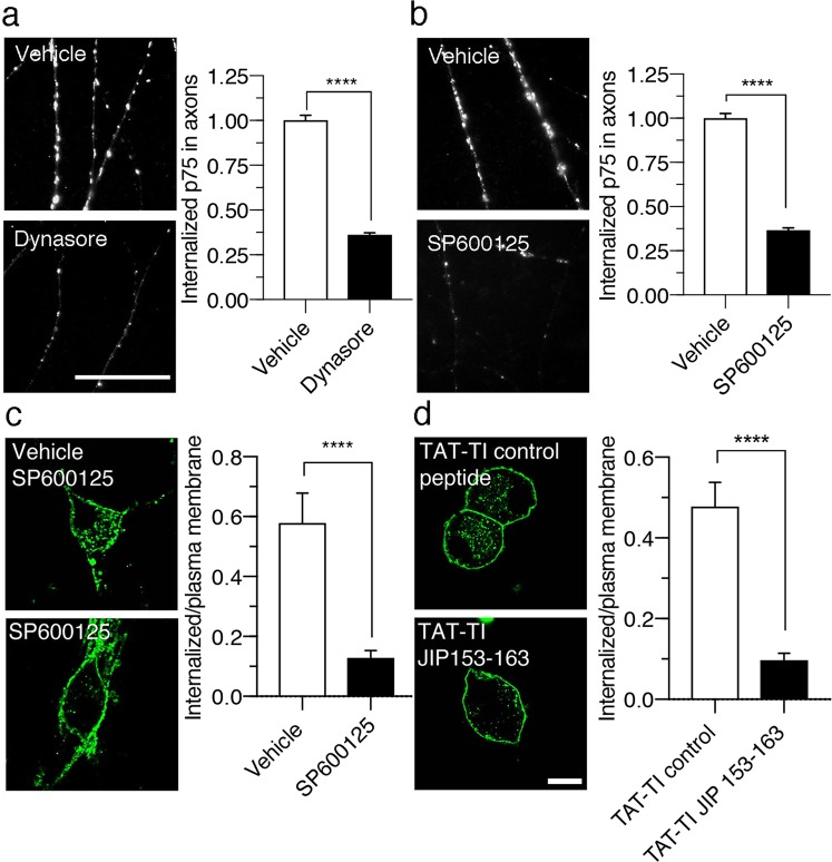
Figure 7.
JNK promotes p75 internalization in the axons and cell bodies of sympathetic neurons. (a) and (b) Visualization of axons of compartmentalized cultures of sympathetic neurons treated with BDNF and MC192-pHRodo in the absence (vehicle) or presence of dynasore for 3–4 hours at 37 °C (a), or the absence (vehicle) or presence of SP600125 for 3–4 hours at 37 °C (b). The intensity of the pHRodo fluorophore is brighter at an acidic pH, indicating p75 internalization. An inhibitor of p75 internalization, dynasore, reduced the fluorescence associated with p75 internalization. Similar to the effect of dynasore, the presence of SP600125 reduces the fluorescence compared to vehicle conditions. Scale bar, 4,5 µm. Right panels show the quantification of the total fluorescence associated with 366 (vehicle for dynasore), 406 (dynasore), 268 (vehicle for SP600125), 227 (SP600125) axonal segments (9 μm long) from three independent compartmentalized neuronal cultures. Statistically significant differences were analyzed using a two-tailed Mann-Whitney test. ****p < 0,0001. (c) and (d) Confocal microscopy images of sympathetic neurons treated with BDNF (150 µg/mL) and MC192-Alexa Fluor 594 (3 μg/mL, green, to label p75) for 4 hours at 37 °C in the absence (vehicle and TAT-TI control peptide) or presence of the JNK inhibitors, SP600125 (10 µM) (c) or TAT-TI-JIP 153–163 peptide (1 μM) (d). Scale bar, 10 μm. Right panels show the levels of internalized p75 after different treatments (relative fluorescence normalized to cell surface p75). Sixty-five cells from three independent experiments were quantified. Statistically significant differences were analyzed using a two-tailed Mann-Whitney test. ****p < 0,0001.

