Figure 1.
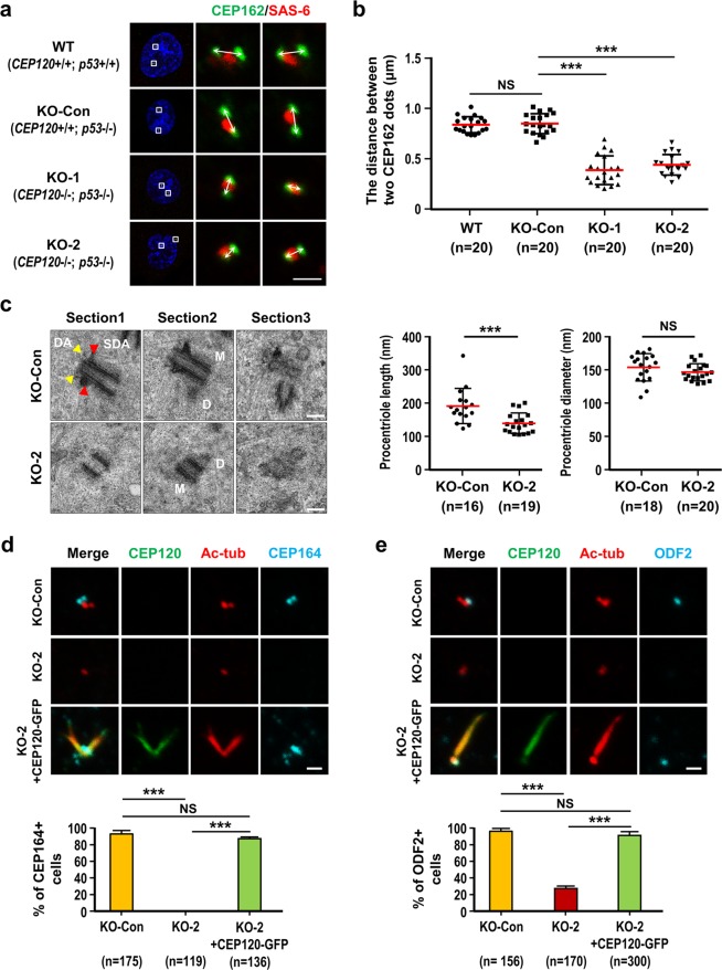
Loss of CEP120 produces short centrioles with no apparent distal appendage (DA) and subdistal appendage (SDA) structures. RPE1-based wild-type cells (WT; CEP120+/+; p53+/+), control cells (KO-Con; CEP120+/+; p53−/−), and knockout cell lines KO-1 (CEP120−/−; p53−/−, clone #3-6-3) and KO-2 (CEP120−/−; p53−/−, clone #34) were treated with aphidicolin for 24 h and released in fresh medium for another 15 h to allow progression to G2 phase. (a) Cells were examined by confocal immunofluorescence microscopy using the indicated antibodies. (b) Histogram illustrating the distances between two CEP162 dots. (c) Representative images of three serial electron microscopy (EM) sections (100 nm/each) of the same centrioles in KO-Con and KO-2 cells. The distal appendages (DA, yellow arrowheads) and subdistal appendages (SDA, red arrowheads) are seen in the mother centriole of KO-Con cells but are absent from KO-2 cells (M: mother; D: daughter). Histogram illustrating the length and diameter of procentrioles in KO-Con and KO-2 cells, as analyzed by EM. Error bars in (b,c) represent the mean ± s.d.; ***P < 0.001; NS, not significant. (d,e) Rescue experiments. KO-Con, KO-2, and KO-2 cells expressing doxycycline-inducible CEP120-GFP WT cells were synchronized at G2, fixed, and stained with the indicated antibodies. Histogram illustrating the percentages of CEP164-positive cells (d, lower panel) or ODF2-positive cells (e, lower panel). Error bars represent the mean ± s.d. from pools of cells (n) from three independent experiments. ***P < 0.001; NS, not significant. Note that overexpression of CEP120-GFP induces centriole over-elongation. Scale bars, 1 μm in (a,d,e) 100 nm in (c).

