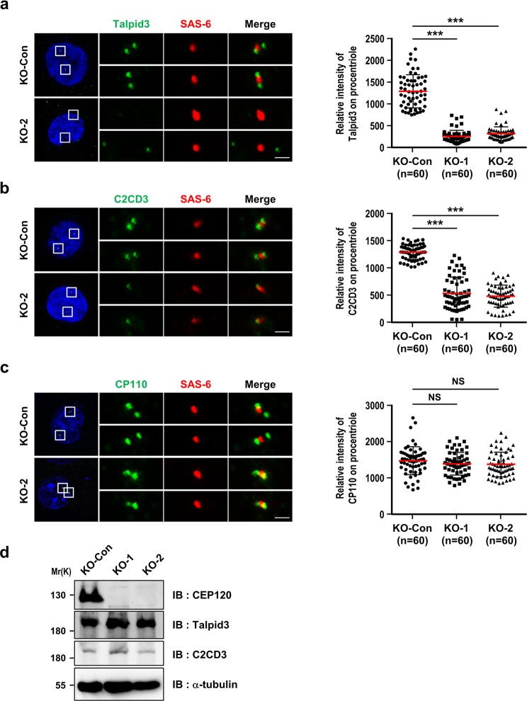
Figure 3.
CEP120 loss shows defective recruitment of the centriolar distal-end proteins C2CD3 and Talpid3. (a–c) KO-Con, KO-1, and KO-2 cells were synchronized at G2 phase and analyzed by immunofluorescence confocal microscopy using antibodies against Talpid3 (a), C2CD3 (b), CP110 (c), and SAS-6 (a–c), and the results were quantified. Histogram illustrating the relative intensity of Talpid3 (a), C2CD3 (b), and CP110 (c) on procentrioles. (d) The protein expression levels of endogenous CEP120, Talpid3 and C2CD3 were examined by immunoblotting using the indicated antibodies. Uncropped blots are shown in Fig. S6a. Error bars represent the mean ± s.d. ***P < 0.001; NS, not significant. Scale bar, 1 μm.

