Figure 6.
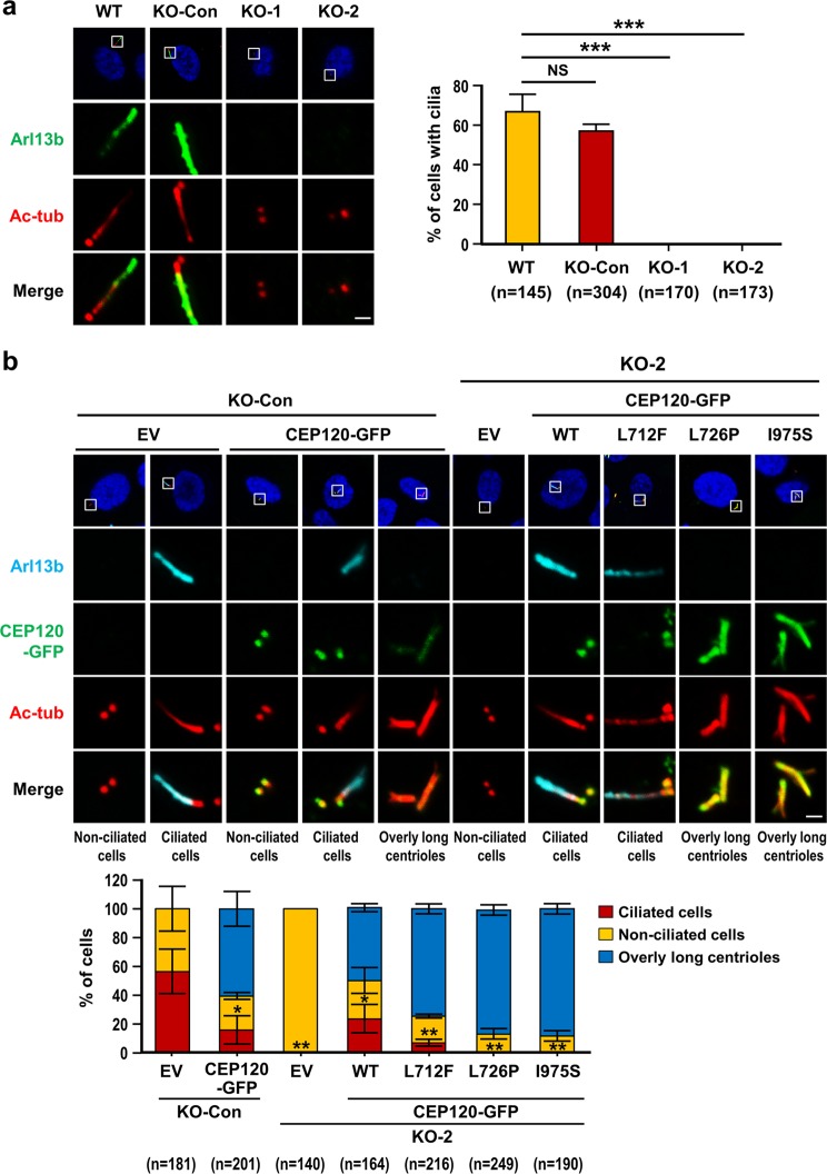
Various disease-associated CEP120 mutants impair cilia formation to different degrees. (a) Loss of CEP120 completely blocks cilia formation. RPE1-based WT (p53+/+; CEP120+/+), KO-Con (p53−/−; CEP120+/+), KO-1 (p53−/−; CEP120−/−), and KO-2 (p53−/−; CEP120−/−) cells were examined by immunofluorescence confocal microscopy using antibodies against Arl13b (a cilium marker, green) and acetyl-tubulin (Ac-tub, red). Histogram illustrating the percentages of cilia-containing cells. Error bars represent the mean ± s.d. from pools of cells (n) from three independent experiments. ***P < 0.001; NS, not significant. (b) Rescue experiments. KO-Con, KO-2, and KO-2 cells expressing doxycycline-inducible WT or mutant CEP120-GFP proteins were serum starved for 48 h to induce cilia formation. Ciliated cells (marked with long Arl13b/Ac-tub filaments), non-ciliated cells (Arl13b-negative), and cells with overly long centrioles (marked with long CEP120-GFP/Ac-tub filaments of >0.5 μm) were examined by confocal fluorescence microscopy using the indicated antibodies. Histogram illustrating the percentages of ciliated cells, non-ciliated cells, and cells with overly long centrioles. Error bars represent the mean ± s.d. from pools of cells (n) from three independent experiments. *P < 0.05; **P < 0.01. Note that CEP120-GFP overexpression induces overly long centrioles (>0.5 μm) that perturb cilia formation in the rescue experiments. EV: empty vector. Scale bar, 1 μm.

