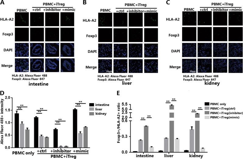
Fig. 8. miR-142-3p affects the infiltration of in vitro induced Tregs (iTregs) and inflammatory cells in different tissues of the xenogeneic graft-versus-host disease (xGVHD) model.
The paraffin-embedded specimens of mouse organs (intestine, liver, kidney) from each xGVHD group on day 21 were detected using immunofluorescence and the human monoclonal antibodies against HLA-A2 or forkhead box P3 (Foxp3) for each group; the secondary antibodies were Alexa Fluor 488 and Alexa Fluor 647. The HLA-A2+ cells and Foxp3+ cells were then measured. The positive fluorescence intensity was measured using the ImagJ software. Representative example of immunofluorescence detection results for a intestine, b liver, and c kidney. d Positive Alexa Fluor 488 fluorescence intensity of specimens from each group indicating HLA-A2+ proportion. e Foxp3+/ HLA-A2+ ratio of specimens from each group. Results are presented as mean ± standard error of the mean values. **P < 0.01. The results shown are representative of two independent xGVHD experiments

