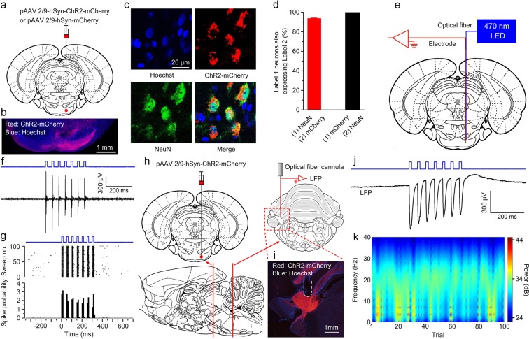
Figure 1.
Selective labelling the right PN neurons and optogenetic stimulation of mossy fibers in the left MCP. (a) The rats were stereotactically injected with pAAV 2/9-hSyn-ChR2-mCherry or pAAV 2/9-hSyn-mCherry targeting the right PN. (b) Example of ChR2-mCherry expression in the right PN. (c) Representative images showing cell-specific ChR2-mCherry expression (red) in neurons (green) of the PN. (d) Statistics of expression in the neurons (468 cells, from 4 mice). (e) In vivo right PN “optrode” recording setup. (f,g) Multi-unit activity in the right PN from a rat injected with pAAV2/9-hSyn-ChR2-mCherry in response to trains of 7 light pulses (470 nm, 10 mW/mm2, 20 Hz, 15 ms pulse duration). Blue bars represent light on. (h) Schematic illustration of in vivo optical stimulation and LFP recording in the left MCP. (i) Example of ChR2-mCherry expression in the left MCP. White dashed line: optrode position. (j,k) Trains of 7 light pulses (470 nm, 25 mW/mm2, 20 Hz, 15 ms pulse duration) also evoked robust LFP responses in the left MCP of a wake behaving rats. Note that the graph. (j) Illustrates an example of the mean value of 100 light-induced LFPs. Data are represented as mean ± s.e.m.

