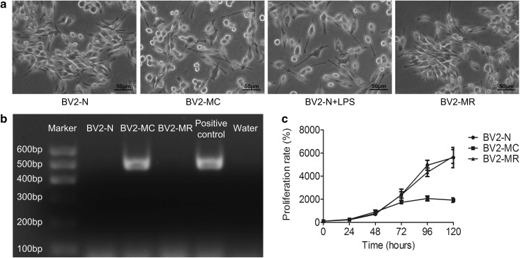
Fig. 1.
Cell morphology and proliferative capability of mycoplasma-contaminated BV2 cells and PCR detection of mycoplasma. a Cell morphology of BV2-N, BV2-MC, LPS-treated BV2-N (BV2-N + LPS) and BV2-MR. b PCR detection of mycoplasma showed that BV2-MC had one specific band at the position of 502-520 bp, similar with positive control. c BV2-MC had a decreased proliferative capability compared to BV2-N. The proliferation of BV2-MC greatly decreased in BV2-MC but restored after mycoplasma elimination. (BV2-N: normal BV2 cell, BV2-MC: mycoplasma-contaminated BV2 cell, BV2-MR: mycoplasma-removed BV2-MC cell) (bar = 50 μm)

