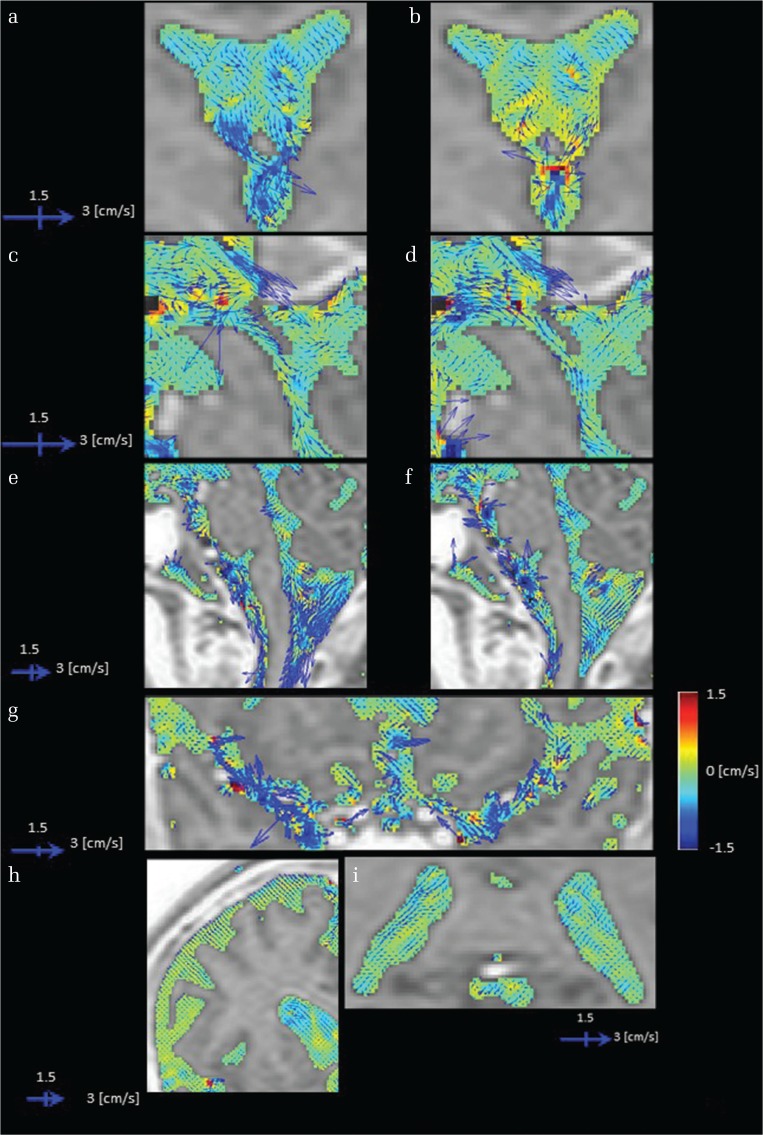
Fig. 1.
Images of cerebrospinal fluid (CSF) velocity with 3DPC in a healthy volunteer. CSF velocity in each imaging plane is represented by the vector length and direction, while the velocity orthogonal to the imaging plane is shown by color coding. The vectors and color scales are as displayed. Sites with irregular motion show long vectors that point in various directions with colors that also change. In addition, blood flow-derived velocity components (non-CSF components) arising from inside the blood vessels were subtracted and removed from the images. Rotational motion within the anterior horn as a vector (a), and the antero-posterior direction as changes on a color scale (b). A motion that ascends toward the lateral and the third ventricle (b and d) and conversely descends toward the third and fourth ventricle (a and c). Irregular motions are evident in the third ventricle based on rotational motion expressed as long vectors and the large change in color display that represents motion orthogonal to the imaging plane (a–d). In the fourth ventricle, augmented motion is observed (e and f). A gentle motion is observed in the trigone (i). A strong motion is shown to strengthen toward the subarachnoid space of the upper cervical spine (e and f). An active CSF motion proximal to the Sylvian fissure that attenuates as it transmits laterally (g), although motion is gradually attenuated toward the distal Sylvian fissure. An augmented motion in a limited area near the vascular structure (g). A suppressed motion seen in convexity (h).

