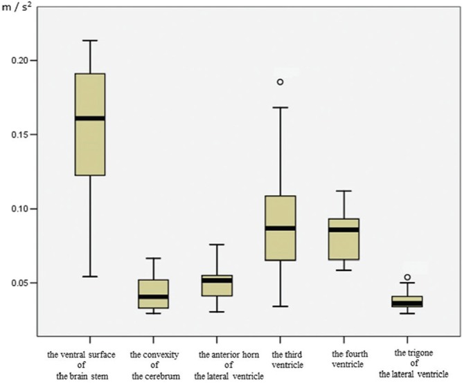
Fig. 4.

Quantitative value of CSF acceleration in the ventricular system and subarachnoid space of healthy volunteers. Healthy volunteers (age range 28–73 years, male = 6, female = 6). Acceleration of CSF is shown as the rate of velocity change per unit time. From a fluid mechanics perspective, sites with small volumes, such as the subarachnoid space at the ventral surface of the brainstem, are considered to have increased fluid velocity compared with sites with large volumes, such as the trigone; however, because acceleration is less affected than velocity by the volume of the site in which the fluid is present, it is excellent for fluid mechanics analysis that compares sites with different volumes. Circles indicate outliers. Reprinted from Takizawa et al.56) (Fig. 4). The quantitative value indicating augmented acceleration in the anterior horn is increased compared with that of the trigone. High CSF acceleration in the third and fourth ventricles. CSF motion demonstrates a gentle CSF acceleration at the trigone compared with other ventricles, consistent with results from other imaging (Figs. 1–3) evaluations. CSF acceleration corroborates the finding that CSF motion in the subarachnoid space from the ventral surface of the brainstem is elevated, whereas acceleration at the convexity of the cerebrum is suppressed.
