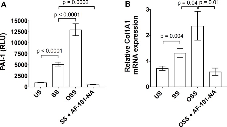
Figure 5.
OSS-activated TGF-β1 stimulated PAI-1 luciferase activity in MLEC and collagen expression in endothelial cells. (A) MLECs were stimulated with OSS or SS or unsheared (US) platelet releasates for 18 hours with and without anti-TGF-β1 neutralizing antibody (AF-101-NA). PAI-1 luciferase activity was measured using luminometer and data were plotted as relative luminescence unit (RLU). (B) HUVECs were stimulated with OSS- or SS-activated platelet releasates or unsheared platelet releasates (US) with and without anti-TGF-β1 antibody for 6 hours, collagen (Col1a1) gene expression was measured by RT-PCR (n = 3–4).

