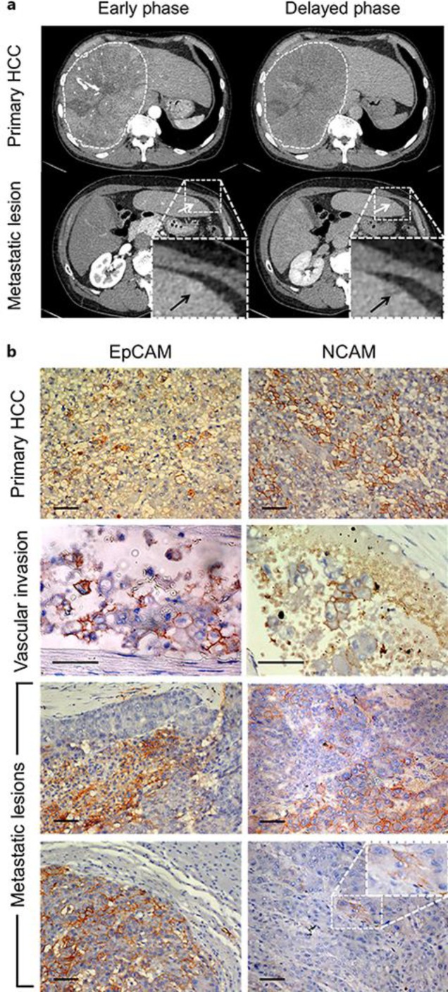
Fig. 1.
Hepatocellular carcinoma (HCC) before operation. a Contrast-enhanced computed tomography of the HCC. A huge HCC occupied most of the right lobe of the liver (dashed white lines circle the HCC). Small metastatic lesions (white and black arrows) were suspected at the surface of the left lobe of the liver. b Histochemical analysis of EpCAM and NCAM expression in the primary and metastatic tumor lesions and in the blood vessels. The tissues were counterstained with hematoxylin. In the primary lesions, 5–10% of cancer cells were EpCAM positive and 10–20% of cancer cells were NCAM positive (uppermost panels). In the blood vessels, cancer cells that were heterogeneously stained for EpCAM and NCAM were frequently detected (panels second from the top). In the metastatic lesions (bottom four panels), heterogeneously stained HPC marker-positive tumors were also detected; however, the frequency of HPC-positive cancer cells differed according to the tumor. Scale bar = 50 μm.

