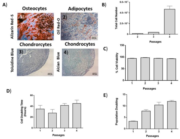
Figure 3.
Differentiation potential, growth kinetics and cell viability of ADMSCs. (A) ADMSCs were successfully differentiated to “osteocytes”, “adipocytes”, and “chondrocytes”. Scale bars 100 μm. (B) Total cell number, (C) cell viability, (D) cell doubling time (CDT), and (E) population doubling (PD) of ADMSCs.

