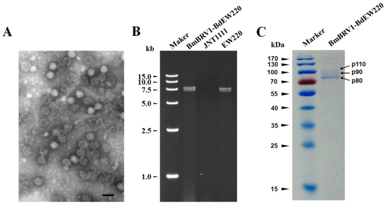
Figure 5.
Features of the BmBRV1-BdEW220 viral particles (A) Transmission electron microscopy (TEM) images of the BmBRV1-BdEW220 viral particles; (B) agarose gel electrophoresis of the dsRNAs extracted from purified virus particles of BmBRV1-BdEW220 and the mycelia of strain EW220 (line EW220); (C) SDS-PAGE analysis of the purified viral particles shows the three distinct protein bands. The scale bar represents 50 nm.

