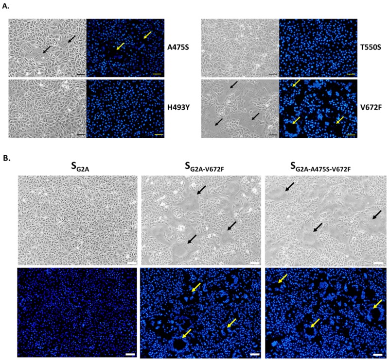
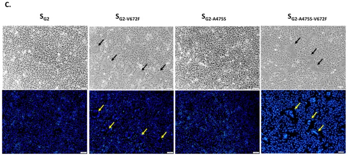
Figure 5.
Effect of amino acid substitution in the RBD of SG2 on syncytium formation in VeroE6-APN cells. (A) VeroE6-APN cells were transfected with pCAGGS expressing SG2A with single amino acid substitution at positions 475, 493, 550, and 672 and treated with trypsin. At 24 hpt, cells were assessed for syncytium formation. Scale 50 µm. (B) VeroE6-APN cells were transfected with pCAGGS expressing SG2A, SG2A-V672F, and SG2A-A475S-V672F and cultured in the presence of trypsin. At 24 hpt, cells were evaluated for syncytium formation. Hoechst was used to stain nuclei. Arrows denote the formation of the syncytium. Scale 100 µm. (C) VeroE6-APN cells were transfected with pCAGGS expressing wild-type SG2, SG2-V672F, SG2-A475S, and SG2-A475S-V672F and cultured in the presence of trypsin. At 24 hpt, cells were evaluated for syncytium formation. Hoechst was used to stain nuclei. Arrows denote the formation of the syncytium. Scale 100 µm.


