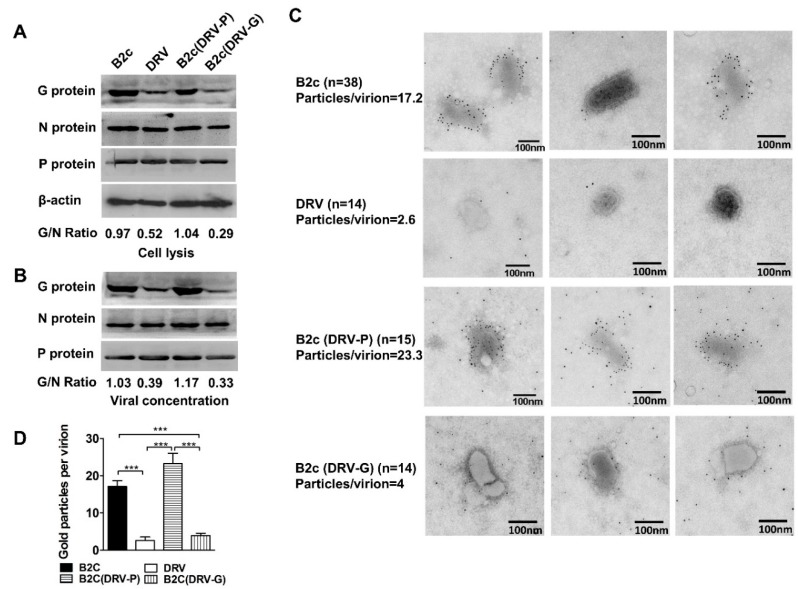
Figure 1.
Expression of RABV-G in NA cells and G incorporation into virions. (A) Western blot of viral protein expression in NA cells infected with indicated RABVs at MOI 1. (B) Western blot analysis of viral protein incorporated into virions. The ratio between G and N was calculated from the intensity of the band using Image-Pro Plus 6.0. (C) Electron micrographs of the indicated RABV virions purified from the supernatants of infected NA cells. RABV-G protein was detected by staining with RABV-G-specific antibodies labeled with 5-nm immunogold particles. (D) The number of gold particles per virion was calculated by manual counting of positive gold dots. Significance of differences between the gold particles per virion of the indicated virus was assessed by the unpaired t-test. *, p < 0.05; **, p < 0.01; ***, p < 0.001.

