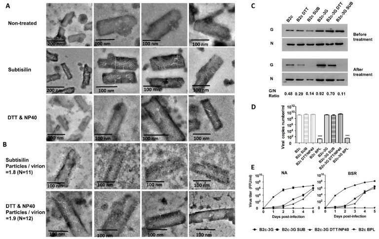
Figure 3.
Removal of G from RABV virions after treatment with DTT/NP40 or subtilisin. (A) Morphological analysis of DTT/NP40- or subtilisin-treated RABV by electron microscopy after negative staining. (B) RABV-G protein on virions was detected by staining with RABV-G-specific antibodies labeled with 5-nm immunogold particles. (C) Western blot analysis of viral N and G proteins incorporated into virions. (D) Viral genomic RNA from RABV treated with or without DTT/NP40 or subtilisin and analyzed by qRT-PCR. (E) Growth curves of B2c-3G and B2c in the NA and BSR cells.

