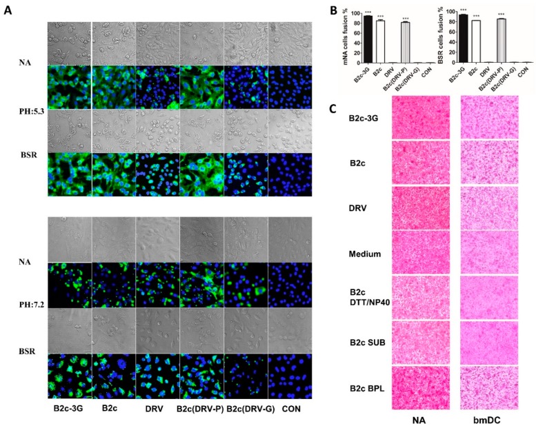
Figure 4.
RABV-G-mediated cell-to-cell fusion. (A) BSR and NA cells were infected with the indicated viruses for 48 h followed by microscopic evaluation of cell-to-cell fusion at pH 7.2 or pH 5.3. (B) Cell fusion index was determined by calculating the ratio of the number of nuclei of multinucleated cells to the number of total nuclei in the photographs taken of randomly selected fields, in which 300 nuclei or more were counted. (C) NA and bmDC cells were treated with the indicated viruses for 4 h before microscopic evaluation of cell-to-cell fusion at pH 5.3.

