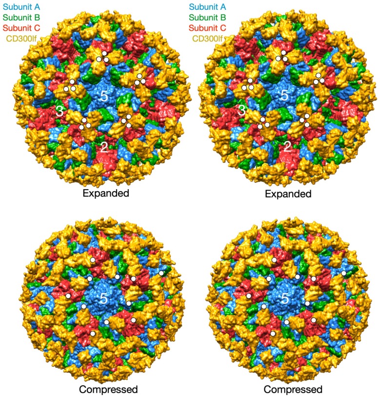
Figure 4.
Models of the bound receptor in the compressed and expanded virions. The structures of the MNV/receptor complex in the expanded state using the cryo-EM structure and the compressed state using the relative P domain orientation observed in NV. The A, B, and C subunits are blue, green, and red respectively and the receptor is shown in yellow. The C-termini of several copies of CD300lf are highlighted by white circles.

