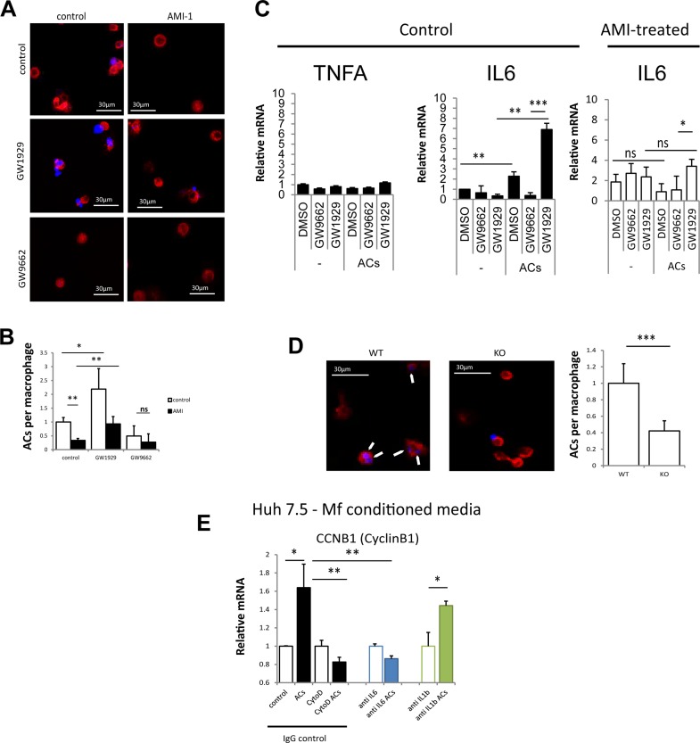
Figure 5.
Macrophage polarization by apoptotic cells requires PRMT1 and PPARγ. (A) THP-1 cells were differentiated into macrophage in the presence or absence of PRMT1 inhibitor AMI-1. Macrophages labeled with phalloidin-568 (red) were treated with apoptotic cells (UV-irradiated hepatocytes) labeled with DAPI (blue) for 1 h in the presence of PPARγ agonist GW1929 or PPARγ antagonist GW9662 where indicated. Scale bars: 30 μm. (B) Average number of phagocytosed apoptotic cells (ACs) per macrophage as in (A) in n = 3 independent experiments. Data presented as average ± SD. *p < 0.05; **p < 0.01. (C) THP-1 monocytes as in (A). Relative mRNA levels after 24 h of ACs in n = 3 independent experiments. Data presented as average ± SD. *p < 0.05; **p < 0.01; ***p < 0.001. (D) Efferocytosis assay as in (A) using isolated macrophages from WT or MKO mice. Arrows indicate internalized apoptotic cells. Scale bars: 30 μm. ***p < 0.001. (E) THP-1 cells were treated with ACs as in (A) in the presence or absence of cytochalasin D. Conditioned media was added to Huh 7.5 cells in the presence of anti-IL-6, anti-IL-1β, or control IgG. Relative mRNA levels of cyclin B1 in n = 3 independent experiments. Data presented as average ± SD *p < 0.05; **p < 0.01.

