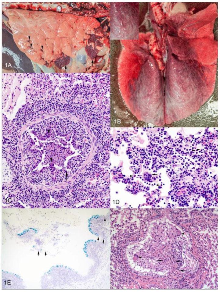
Figure 1.
Gross and microscopic pathology of experimental BRSV infection. (A) Lung from calf experimentally infected with bRSV and examined 7 days later. Ventral areas of right cranial and right middle lung lobes are atelectic and dark red-purple (arrows). Similar small lesions are visible in the right caudal lobe (arrowheads). The remainder of the lung failed to collapse. (B) Lung from calf experimentally infected with bRSV and examined 7 days later. There is an overall red discoloration to the lungs. Dorsal regions of all lobes are characterized by interlobular and subpleural edema (inset). (C) Photomicrograph of lung from calf experimentally infected with bRSV and examined 7 days later. There is degeneration and necrosis of bronchial epithelium with sloughed cells and debris in bronchial lumen. Some areas lack epithelium (arrows). Note epithelial syncytia (arrowheads). (D) Photomicrograph of lung from calf experimentally infected with bRSV and examined 7 days later. Interstitial capillaries are congested and alveolar interstitium contains numerous mononuclear cells. Alveolar lumina contain macrophages, neutrophils (arrowheads) and syncytia (arrow). (E) Photomicrograph of lung from calf experimentally infected with bRSV and examined 7 days later. In situ hybridization using probes for bRSV F protein (green) and IL-8 (red). (F) Photomicrograph of lung from calf experimentally infected with bRSV and examined 14 days later. Bronchiolitis obliterans; polypoid proliferative epithelium, including syncytia (arrowheads), supported by a fibrous stalk (arrows) fills bronchiolar lumen.

