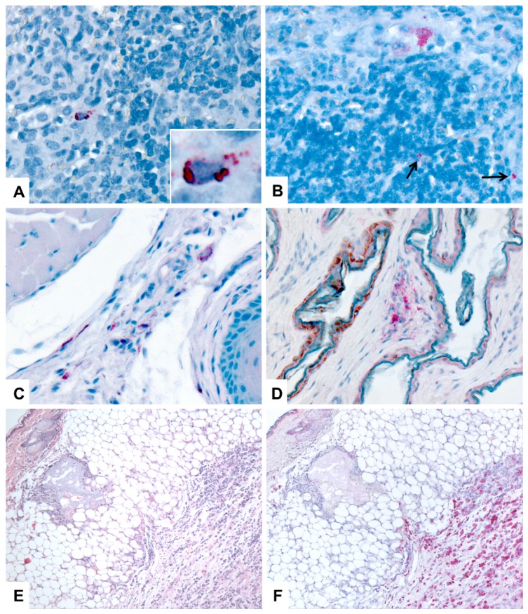
Figure 5.
Immunohistochemical (IHC) localization of Marburg viral (MARV) antigen in tissues of Egyptian rousette bats. All IHC stains are immunoalkaline phosphatase with naphthol fast red and hematoxylin counterstain. DPI = days post-inoculation. (A) Spleen, 5 DPI. MARV antigen (red) is present in the cytoplasm of small numbers of red pulp histiocytes. Original magnification: 400x. Inset: higher magnification of a histiocyte showing granular to globular, cytoplasmic staining of antigen. Original magnification: 1000×. (B) Axillary lymph node; 10 DPI. Marburg viral antigen is localized to the cytoplasm of histiocytes in the subcapsular sinus (top of image) and in the paracortical region (arrows). Original magnification: 400×. (C) Tongue (mucosa, submucosa, and skeletal muscle); 9 DPI. Marburg virus antigen (red) is present in a small number of histiocytes and fibroblast-type cells. Original magnification: 400x. (D) Skin, patagium (wing membrane), 10 DPI. Cytoplasmic antigen (red) is present in a focus of dermal histiocytes. Original magnification: 400×. (E) Skin and subcutaneous tissue from the MARV inoculation site, 3 DPI. The subcutis is infiltrated by a dense aggregate of macrophages at the site of viral inoculation. HE stain; original magnification: 50×. (F) Skin and subcutaneous tissue from the MARV inoculation site (replicate of section in C). Immunohistochemical stain demonstrating Marburg viral antigen (red) in macrophages in the subcutaneous tissues. Immunoalkaline phosphatase stain with naphthol fast red and hematoxylin counterstain; original magnification, 50×.

