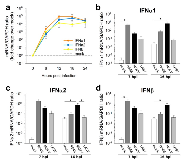
Figure 2.
IFN-I production in LASV-infected pDCs is less long-lasting than that of MOPV-infected pDCs. (a) pDCs were infected with MOPV (MOI = 2). Every 6 h, from 0 to 24 hpi, IFN-I mRNA was quantified by RT-qPCR. Data are presented as the fold change in the mRNA/GAPDH ratio in MOPV-infected pDCs relative to uninfected pDCs. (b–d) pDCs were cultured for 7 h or 16 h in culture medium (mock), R848 (1 µg/mL), MOPV, or LASV (MOI = 2). IFNα1 (b), IFNα2 (c) and IFNβ (d) mRNAs were quantified by RT-qPCR. Data shown are the means and SEM of three (a), four (b–d – 7 hpi), or seven (b–d – 16 hpi) independent experiments. ANOVA on Ranks followed by pairwise comparisons (Tukey test) were performed. Differences are significant for p < 0.05. Significant pairwise comparisons are indicated by a star (*).

