Figure 3.
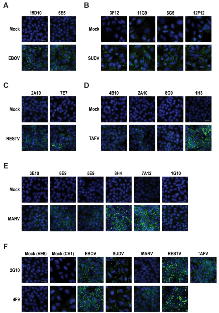
Anti-NP filovirus mAbs detected the native filovirus NP in confocal microscopy (CM). In CM, the cells were infected with a multiplicity of infection (MOI) of 3, fixed and permeabilized at 48 h post-infection (hpi), and stained with different anti-filovirus NP mAbs; the nuclei were stained with DAPI, and finally filovirus NP expressions were detected by treating with Alexa 488 secondary anti-mouse antibodies (Abs). Mock infection controls were also maintained with both primary and secondary Ab staining. (A) Vero E6 cells were infected with EBOV treated with 15D10 and 6E5 mAbs. (B) CV1 cells were infected with SUDV and treated with 3F12, 11G9, 6G5, and 12F12 mAbs. (C) Vero E6 cells were infected with RESTV and stained with primary 2A10 and 7E7 mAbs. (D) Vero E6 cells were infected with TAFV and treated with 4B10, 2A10, 8G9, and 1H3 mAbs. (E) Vero E6 were infected with MARV and treated with 3E10, 6E9, 5E9, 8H4, 7A12, and 1G10 mAbs. (F) CV1 cells were infected with SUDV, and Vero E6 cells were infected with EBOV, MARV, RESTV, and TAFV; therefore, they were treated separately with 2G10 and 4F9 to check cross reactivities against different filoviruses. The scale bar represents 20 µm.

