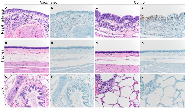
Figure 5.
Histopathological changes in the respiratory tract of vaccinated and unvaccinated alpaca after challenge with MERS-CoV. Nasal turbinate, trachea, and lung were collected from vaccinated (A–F; A1 shown) and unvaccinated control animals (G–L; A3 shown) on 5 dpi and stained with hematoxylin and eosin (A–C and G–I), or a polyclonal anti-MERS-CoV antibody panels (D–F and J–L). MERS-CoV antigen is visible as a red brown staining in the immunohistochemistry panel. No histopathological changes were observed in the vaccinated alpaca whereas the unvaccinated control alpaca displayed multifocal, minimal-to-mild, acute rhinitis. MERS-CoV antigen was only detected in the nasal turbinates. Magnification: 400×.

