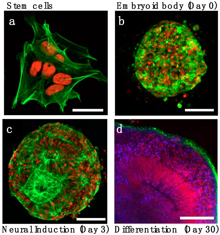
Figure 1.
Stages of in vitro organoid development. (a) Stem cells with live reporters H2B-mCherry (red) and lifeact-GFP (green) are assembled into (b) 3D embryoid bodies. (c) Following neural induction, ventricle-like lumens appear within the 3D culture. (d) Neural differentiation and layer organization with neuronal progenitor (PAX6+, red) surrounding the lumen, an intermediate layer of sparse progenitors (DAPI, blue) and neuronal layer (TUJ1+, green) on the organoid periphery. Images adapted from Karzbrun et al. [7]. Scale bars are (a) 20 μm and (b–d) 100 μm.

